

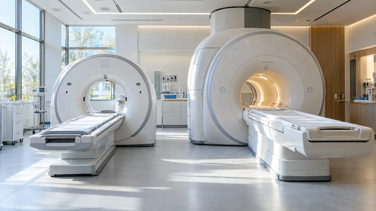
Computed tomography (CT) scans create images of internal organs using X-rays, while magnetic resonance imaging (MRI) uses a strong magnetic field and radio waves without producing ionizing radiation. In practice, CT is faster and better suited for emergency situations, as well as for assessing bone and lung health, while MRI provides more detailed images of soft tissues: the brain, spinal cord, joints, and ligaments. The doctor always decides which test to perform based on the patient's symptoms and the specific investigation they wish to perform.
To sum it up
- Computed tomography uses x-rays, resonance – magnetic fields and radio waves.
- A CT scan typically takes a few minutes, while an MRI scan takes anywhere from a few minutes to an hour, depending on the area being examined.
- CT is the method of choice for injuries and emergency conditions (for example, if intracerebral hemorrhage is suspected), MRI is the method of choice for diseases of the brain, spinal cord, joints and ligaments.
- Contraindications to MRI may include metal elements and devices in the body (for example, some pacemakers), and to CT - pregnancy and allergy to iodine-containing contrast.
Computed Tomography and Magnetic Resonance Imaging – How Do They Work?
Computed tomography — is an advanced version of traditional radiography. An X-ray tube rotates around the patient, and detectors measure the amount of radiation absorbed by individual tissues. A computer combines these measurements into cross-sectional images of the body. Structures of varying densities—bones, air in the lungs, fresh blood—are clearly visible.
MRI It works differently. A strong magnetic field aligns hydrogen protons in tissues (primarily in water and fat), and radio wave pulses briefly excite them. When the protons return to their original state, they emit a signal that creates an image. Because tissues vary in water content, MRI allows for excellent resolution of soft tissues—the gray and white matter of the brain, cartilage, ligaments, and intervertebral discs.
Externally, these machines are similar: the patient lies on a table that slides into the machine's opening. However, a CT scanner has a short, wide ring, while an MRI typically has a longer, narrower bore.
Comparison Chart: CT Scan vs. MRI
| Characteristic | Computed tomography (CT) | Magnetic resonance imaging (MRI) |
|---|---|---|
| Operating principle | X-ray radiation | Magnetic field and radio waves |
| Ionizing radiation | Yes | NO |
| The time of the exam itself | Usually from a few seconds to a few minutes. | Usually 20-60 minutes |
| This shows it best. | Bones, lungs, bleeding, trauma | Brain, spinal cord, joints, ligaments, soft tissues |
| Contrast | Iodine | gadolinium |
| Noise | Small | Loud knock - the patient is given earplugs or headphones. |
| Main contraindications | Pregnancy, allergy to iodine-containing contrast, renal failure (if contrast is present) | Some metal implants and electronic devices in the body |
When should CT and MRI be used? Areas of application.
Computed tomography (CT) is fast and widely available This is a diagnostic method, so hospital emergency departments use it first. Typical indications include:
- Head, spine and multiple organ injuries (eg following an accident),
- If a stroke is suspected, CT allows for rapid differentiation between bleeding and ischemia.
- diseases of the lungs and chest,
- complex fractures,
- Diagnosis of the abdominal cavity and pelvis, assessment of blood vessels (angio-CT).
MRI more accurate for soft tissue examination . Doctors refer patients for this procedure, in particular:
- diseases of the brain and spinal cord (eg, multiple sclerosis, tumors),
- discopathy and pressure on the nerves in the spine.
- Joint injuries – knee, shoulder, ligaments and meniscus.
- accurate assessment of changes in the abdominal and pelvic organs.
Therefore, the question "Which is better—CT or MRI?" cannot be answered without examining the underlying cause. With head injuries, every minute counts, and CT is preferable, whereas with chronic knee pain, MRI will provide more complete information. These two methods often complement each other.
Safety and contraindications
Computed tomography
Computed tomography (CT) scan involves exposure to ionizing radiation , so it is only performed when the diagnostic benefit outweighs the risk. Particular caution should be exercised when examining children, and CT scans are generally not performed on pregnant women. For examinations using iodinated contrast agents, previous allergic reactions to the contrast, kidney health, and the presence of thyroid disease are important—so a questionnaire is completed before the examination, and a creatinine test is often ordered.
Magnetic resonance imaging
MRI does not use ionizing radiation, but its strong magnetic field can be dangerous for people with metal objects or devices in the body : pacemakers and other stimulators, cochlear implants, some vascular clamps, or metal fragments. Many modern implants are approved for MRI under certain conditions, but the laboratory makes the decision based on the implant documentation. MRI can be performed during pregnancy, but is not recommended in the first trimester, and gadolinium contrast is administered only in justified cases.
Examination progress and patient comfort
Both tests require you to lie still and remove all metal objects. However, there are noticeable differences. Computer tomography short and quiet, and the camera ring only covers part of the body, so it rarely causes disturbance.
MRI is a longer and noisier test The machine makes a rhythmic clicking sound while operating, so patients are given earplugs or headphones. Those with claustrophobia may find the tunnel uncomfortable; it's worth mentioning this at registration, as the lab may offer a solution (such as a doctor-prescribed sedative or a machine with a wider opening). Children who are unable to lie still may undergo the examination under general anesthesia.
How to prepare and where to get a referral to a specialist?
The examination, funded by the National Health Fund, requires doctor's referral , which will determine which area of the body should be examined and whether a contrast agent should be used. Both tests may be performed for a fee in private clinics, but it's still worth getting a referral or consulting—the doctor will choose the appropriate imaging method, and the radiologist will know what to look for. MRI is usually more expensive and less accessible than CT because it takes longer, and the equipment is more expensive to purchase and maintain.
Before a contrast study, you'll usually need to fast for several hours and provide creatinine test results—the clinic will provide detailed instructions when you schedule your appointment. Bring your previous test results, and before the MRI, bring documentation confirming the presence of any implants.
Frequently Asked Questions (FAQ)
Which is more accurate – CT or MRI?
It depends on the organ being examined. MRI more accurately depicts soft tissues (brain, spinal cord, joints, ligaments), while CT scans show bones, lungs, and recent bleeding. The doctor decides which imaging method to use.
Is MRI safer than CT?
An MRI doesn't expose you to ionizing radiation, so the procedure can be repeated more frequently. However, it has its own contraindications, primarily related to metal implants and electronic devices in the body. Before the examination, you must complete a safety questionnaire.
How long do computed tomography (CT) and magnetic resonance imaging (MRI) scans take?
A computed tomography (CT) scan itself typically takes anywhere from a few seconds to several minutes. An MRI scan typically lasts between 20 and 60 minutes, depending on the area and number of scans.
Is it possible to have a CT scan or MRI during pregnancy?
Computed tomography (CT) scans are generally not performed on pregnant women due to radiation exposure. MRI may be performed, but is not recommended in the first trimester, and gadolinium contrast is used only in clearly indicated cases. The decision is made by the attending physician.
Is it possible to have an MRI if you have a pacemaker?
Pacemakers and other pacemakers are classic contraindications for MRI. Some newer devices are approved for MRI under certain conditions, but this requires documentation and laboratory approval. Patients with pacemakers may undergo CT scanning.
See also
- What is the difference between scintigraphy and PET?
- What is the difference between implantation bleeding and menstruation?
- What is the difference between complete and basic morphology?
- What is the difference between hysteroscopy and curettage?