

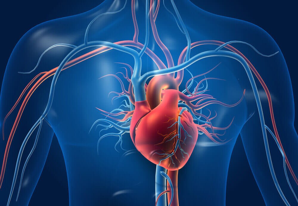
Arteries differ from veins primarily in the direction of blood flow: arteries carry blood from the heart to the organs, and veins carry blood from the organs back to the heart. This leads to further differences: arterial walls are thicker, more muscular and elastic because the blood flows under high pressure, whereas vein walls are thinner and - especially in the extremities - have valves that prevent backflow of blood.
To sum it up
- Direction: Arteries – from the heart, veins – toward the heart. The name does not determine whether the blood is saturated with oxygen.
- Pressure: high and pulsating in the arteries (the pulse is felt here), low in the veins.
- Walls: Arteries have a thick, muscular, and elastic tunica media; veins have a thinner, more flexible tunica media.
- Valves: present in many veins (especially in the lower extremities), but absent from arteries.
The direction of flow is the most important difference.
Many people remember that arteries carry "clean" blood, and veins carry "dirty" blood. This is a simplification that is not always true. The vessel is called artery , when it carries blood away from the heart, and vein , when it delivers it to the heart - regardless of the oxygen content in the blood.
В systemic circulation arteries (led by the aorta) deliver oxygen-rich blood to all organs, and veins collect oxygen-depleted blood and return it to the right atrium. systemic circulation (pulmonary circulation) the situation is reversed: pulmonary arteries deliver oxygen-depleted blood from the right ventricle to the lungs, and pulmonary veins — oxygenated blood into the left atrium. This is a common question on biology exams.
The structure of arteries and veins – walls, lumen and valves.
The wall of an artery and vein consists of three layers: intimacy (lined by endothelium), media (smooth muscles and elastic fibers) and outer membrane , formed by connective tissue. The ratio of these layers varies.
В arteries The intima media is thick and rich in muscle and elastic fibers. This allows the wall to withstand high pressure, stretching with each heartbeat and releasing energy during diastole, thereby regulating blood flow. In cross-section, the artery maintains a round shape.
Vienna They have a thinner middle layer (tunica media), containing less muscle and more connective tissue. Their walls stretch easily, and their lumen is usually wider than that of the accompanying artery. Therefore, veins contain the majority of the body's blood—physiologists call them capacitive vessels . Many veins, especially in the lower extremities, have valves — folds of the inner lining that allow blood to flow only to the heart.
Blood pressure, heart rate, and blood return to the heart
Blood flows through the arteries under pressure created by the contractions of the left ventricle. The wave that propagates along the arterial walls with each contraction is pulse , which we feel, for example, on the wrist (radial artery) or on the neck (carotid artery). In common usage, the term "blood pressure" also refers to arterial pressure.
The pressure in the veins is low and insufficient to push blood from the legs back to the heart. This is facilitated by muscle pump Muscles contracting during exercise compress the veins, and valves ensure that blood flows only upward. This pressure is also maintained by changes in chest pressure during breathing. Therefore, prolonged standing or sitting contributes to leg swelling.
Arteries and veins - comparison table
| Characteristic | Arteries | Vienna |
|---|---|---|
| Flow direction | From the heart to the organs | From organs to the heart |
| Blood (large volume of blood) | Oxidized, light red | Deoxygenated, dark red |
| Exception: pulmonary circulation | Pulmonary arteries: deoxygenated blood | Pulmonary veins: oxygenated blood |
| Pressure | High, pulsating | Short |
| Wall | Dense, muscular, elastic | More subtle, more receptive |
| Valves | Flaw | Present in many veins, especially in the extremities. |
| Position | Mostly deep | Deep and superficial (visible under the skin) |
| Examples of diseases | Atherosclerosis, aneurysm | Varicose veins, deep vein thrombosis |
How to distinguish arterial bleeding from venous bleeding?
This knowledge is useful when providing first aid. Arterial bleeding usually profuse, bright red, and may flow in a pulsating stream that matches the heartbeat. Venous bleeding, usually presents as a constant stream of darker blood. In both cases, the most important thing is to call for help (112 or 999) and apply direct pressure to the wound using a bandage; in case of bleeding from a large vessel, time is of the essence.
Where does the color of the veins visible under the skin come from? Venous blood is not blue, but dark red. The bluish tint is the result of light penetrating the skin and reflecting off the tissue. Blood for analysis Blood is usually taken from veins (for example, from the elbow) because they are located close to the surface and have low pressure. If you notice painful swelling in your calf, dilated veins in your legs, or a weak pulse in your limb, consult a doctor.
Frequently Asked Questions (FAQ)
Do arteries always carry oxygenated blood?
No. The exception is the pulmonary arteries, which carry deoxygenated blood from the right ventricle of the heart to the lungs. The pulmonary veins, on the other hand, carry oxygenated blood to the left atrium.
Why do veins have valves but arteries don't?
In arteries, blood flows under high pressure generated by the heart, preventing it from flowing backward. In veins, the pressure is low, and blood from the lower extremities must overcome gravity—valves prevent backflow.
Where is the pulse measured – in an artery or in a vein?
The pulse is a wave that travels along the walls of the arteries with each heartbeat. It is most often measured on the radial artery at the wrist or on the carotid artery.
Which has a thicker wall: an artery or a vein?
An artery has a thicker tunica media, composed of smooth muscle and elastic fibers, allowing it to withstand high pressure. The vein's wall is thinner and more susceptible to stretching.
How are capillaries different from arteries and veins?
Capillaries connect the smallest arteries with the smallest veins. Their walls consist of almost a single layer of endothelial cells, which ensures the exchange of oxygen, carbon dioxide, and nutrients with the tissues.
See also
- What is the difference between the circulatory and lymphatic systems?
- What is the difference between a stroke and a hemorrhage?
- What is the difference between smooth and striated muscle?
- What is the difference between the abdomen and the stomach?